


Scientists have mapped the intricate details of a small piece of human brain, uncovering mysteries such as nerve cell pairs with unusually tight bonds and axons forming mysterious whorls. The brain sample was taken from a woman during epilepsy surgery and imaged using a high-powered microscope. The goal of brain mapping is to understand how the human brain functions and what goes wrong in various brain diseases.
One surprising finding was that certain nerve cell pairs had more than 50 contact points between them, indicating an exceptionally strong bond. Another discovery was axons forming intricate loops and knots, whose purpose remains unknown. Additionally, mirror-image twin nerve cells were found in the deepest layer of the brain sample, posing yet another mystery. These findings open up new avenues for research into the intricate workings of the human brain.
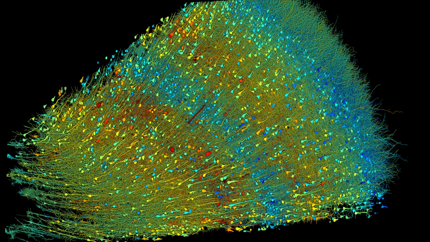
The brain map was created by digitally reconstructing the three-dimensional objects in the brain sample, revealing unprecedented detail about the complex network of cells, synapses, and blood vessels. This level of detail allows scientists to gain new insights into the inner workings of the human brain. Future research aims to scale up the mapping process to larger brain samples and explore similar structures in mouse brains to further understand brain function.
The brain sample used for mapping was obtained with the woman’s consent during epilepsy surgery, emphasizing the importance of ethical considerations in brain research. The exploration of the brain’s landscapes provides a glimpse into the remarkable complexity of the human brain and highlights the need for continued research to unravel its mysteries. Through advancements in technology and computational methods, scientists are uncovering new details about the brain that will shape our understanding of neurological disorders and brain function.


